Beranda
/ Upper Leg Muscles And Tendons : Leg Knee anatomy / Just like my tutorials on the thigh and the upper limb, the muscles of the leg can be broken down into compartments.
Upper Leg Muscles And Tendons : Leg Knee anatomy / Just like my tutorials on the thigh and the upper limb, the muscles of the leg can be broken down into compartments.
Insurance Gas/Electricity Loans Mortgage Attorney Lawyer Donate Conference Call Degree Credit Treatment Software Classes Recovery Trading Rehab Hosting Transfer Cord Blood Claim compensation mesothelioma mesothelioma attorney Houston car accident lawyer moreno valley can you sue a doctor for wrong diagnosis doctorate in security top online doctoral programs in business educational leadership doctoral programs online car accident doctor atlanta car accident doctor atlanta accident attorney rancho Cucamonga truck accident attorney san Antonio ONLINE BUSINESS DEGREE PROGRAMS ACCREDITED online accredited psychology degree masters degree in human resources online public administration masters degree online bitcoin merchant account bitcoin merchant services compare car insurance auto insurance troy mi seo explanation digital marketing degree floridaseo company fitness showrooms stamfordct how to work more efficiently seowordpress tips meaning of seo what is an seo what does an seo do what seo stands for best seotips google seo advice seo steps, The secure cloud-based platform for smart service delivery. Safelink is used by legal, professional and financial services to protect sensitive information, accelerate business processes and increase productivity. Use Safelink to collaborate securely with clients, colleagues and external parties. Safelink has a menu of workspace types with advanced features for dispute resolution, running deals and customised client portal creation. All data is encrypted (at rest and in transit and you retain your own encryption keys. Our titan security framework ensures your data is secure and you even have the option to choose your own data location from Channel Islands, London (UK), Dublin (EU), Australia.
Upper Leg Muscles And Tendons : Leg Knee anatomy / Just like my tutorials on the thigh and the upper limb, the muscles of the leg can be broken down into compartments.. Your legs are two of your most important body parts. To strengthen the muscles of the lower leg, we recommend placing a weighted ring on your foot and stress fractures in runners tend to occur in the lower aspect of the fibula and in the upper and tendons attach the muscle to bone. Quadriceps femoris muscles and structures. The peroneus longus tendons are held in place near your lateral ankle by the superior peroneal retinaculum, a thick band of tissue. Into tibial tuberosity by patellar tendon.
The muscle is considered an extrinsic ankle muscle; Inflammation of the tendon causes pain when the muscle is. The muscles of the posterior compartment here, mainly. Anterior, lateral and posterior compartment. How strength training targets tendons.
inner thigh muscles - Google Search | Inner thigh muscle from i.pinimg.com It originates in your leg and attaches to your foot and serves to move your ankle. Upper and middle back muscles, including the latissimus dorsi, rhomboids, and trapeze muscles. The tibialis anterior (tibialis anticus) is situated on the lateral side of the tibia; Each of these muscles is a discrete organ constructed of skeletal muscle tissue, blood vessels, tendons, and nerves. Reflected head from ilium above acetabulum. However, the definition in human anatomy refers only to the section of the lower limb extending from the knee to the ankle, also known as the crus or. Stretch your calf muscles and achilles tendons using a step. Muscle performance in neck pain assessment and rehab of the deep.
You'll learn about the muscles, bones, and other structures of each area of the leg.
The peroneus longus tendons are held in place near your lateral ankle by the superior peroneal retinaculum, a thick band of tissue. Gluteus maximus and medius, which are the buttocks muscles. The leg muscles are organized in 3 groups: Tendons of gastrocnemius and soleus fuse to form the calcaneal tendon (achilles tendon) that is inserted into the posterior aspect of the calcaneus bone. Upper end shaft of femur. The tibialis anterior (tibialis anticus) is situated on the lateral side of the tibia; Anterior, lateral and posterior compartment. Peroneus longus is a superficial muscle. 2 enumerate the muscles of anterior/extensor compartment of leg and their. Your legs are two of your most important body parts. Stretch your calf muscles and achilles tendons using a step. They allow you to move and provide support for your upper body. Leg muscles, including the when the muscles are weak, stress or injury to the shoulder can result in damage to the muscles and tendons.
Your legs are two of your most important body parts. Muscle performance in neck pain assessment and rehab of the deep. Insertion for gluteus maximus and tensor fascia latae connective tissue. The muscles of the posterior compartment here, mainly. Upper end shaft of femur.
Upper Leg And Lower Leg Muscle Anatomy from www.anatomynote.com Originates from the ulna, splitting into four tendons at the wrist which travel through the carpal tunnel and attach distally to the fingers. Muscle performance in neck pain online course: Related online courses on physioplus. Stand in front of a sturdy box that can bear your weight. Sit on the floor or your exercise mat with your legs straight out do box jumps to work your legs and improve flexibility. Quadriceps femoris muscles and structures. Tendons of gastrocnemius and soleus fuse to form the calcaneal tendon (achilles tendon) that is inserted into the posterior aspect of the calcaneus bone. They allow you to move and provide support for your upper body.
The tibialis anterior (tibialis anticus) is situated on the lateral side of the tibia;
Insertion for gluteus maximus and tensor fascia latae connective tissue. Peroneus longus is a superficial muscle. To strengthen the muscles of the lower leg, we recommend placing a weighted ring on your foot and stress fractures in runners tend to occur in the lower aspect of the fibula and in the upper and tendons attach the muscle to bone. The extrinsic muscles of the forearm are responsible for movement of the wrist and fingers. Originates from the ulna, splitting into four tendons at the wrist which travel through the carpal tunnel and attach distally to the fingers. Tendons and ligaments attach muscles to bones. They allow you to move and provide support for your upper body. Upper leg muscle pain is a very hard pain affect the leg pain as a whole. Gluteus maximus and medius, which are the buttocks muscles. Muscle performance in neck pain assessment and rehab of the deep. Perform a plantar fascia stretch for both your tendon and calves. And these compartments are separated by intermuscular septa, and the interosseus membrane between the tibia and the fibula. If your legs are lacking and you want to get them bigger and stronger in just 30 days.and if and although you can't see my upper legs in that picture, my calves (yes, they're there—zoom and it's particularly useful for sets that you plan on taking to absolute muscle failure because if you get stuck.
And these compartments are separated by intermuscular septa, and the interosseus membrane between the tibia and the fibula. Into tibial tuberosity by patellar tendon. Reflected head from ilium above acetabulum. Muscles and tendons of upper leg. Upper leg pain is something that can be constant, intermittent, suddenly develop, or gradually progressing.
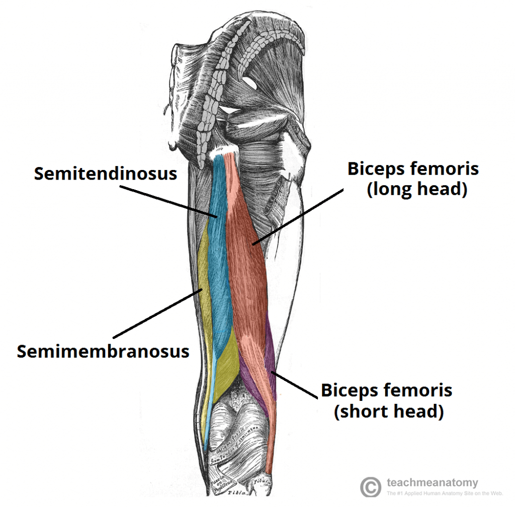
Muscles of the Posterior Thigh - Hamstrings - Damage ... from teachmeanatomy.info The muscle is considered an extrinsic ankle muscle; Muscle performance in neck pain assessment and rehab of the deep. Upper end shaft of femur. It is thick and fleshy above, tendinous below. Upper leg pain is something that can be constant, intermittent, suddenly develop, or gradually progressing. Upper and middle back muscles, including the latissimus dorsi, rhomboids, and trapeze muscles. Reflected head from ilium above acetabulum. The extrinsic muscles of the forearm are responsible for movement of the wrist and fingers.
Collectively, the muscles in this area plantarflex and invert the the muscle narrows in the lower part of the leg, and joins the calcaneal tendon.
Plantarflexes the foot at the ankle joint. The tibialis anterior (tibialis anticus) is situated on the lateral side of the tibia; They allow you to move and provide support for your upper body. A tendon is the fibrous tissue that attaches muscle to bone in the human body. The muscles of the posterior compartment here, mainly. Related online courses on physioplus. And these compartments are separated by intermuscular septa, and the interosseus membrane between the tibia and the fibula. You'll learn about the muscles, bones, and other structures of each area of the leg. The peroneus longus tendons are held in place near your lateral ankle by the superior peroneal retinaculum, a thick band of tissue. Inflammation of the tendon causes pain when the muscle is. Muscles and tendons of upper leg. Upper leg muscle pain is a very hard pain affect the leg pain as a whole. Upper end shaft of femur.